| |
 |
Echo Cardiography (2D, M Mode) |
|
An echocardiogram, often referred to in the medical community as a cardiac ECHO or simply an ECHO, is a sonogram of the heart. Also known as a cardiac ultrasound, it uses standard ultrasound techniques to image two-dimensional slices of the heart.
In addition to creating two-dimensional pictures of the cardiovascular system, an echocardiogram can also produce accurate assessment of the velocity of blood and cardiac tissue at any arbitrary point using pulsed or continuous wave Doppler ultrasound. This allows assessment of cardiac valve areas and function, any abnormal communications between the left and right side of the heart, any leaking of blood through the valves (valvular regurgitation), and calculation of the cardiac output as well as the ejection fraction. |
|
| |
|
| |
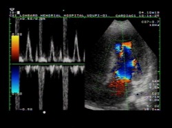 |
COLOR DOPPLER |
|
A Doppler ultrasound test uses reflected sound waves to evaluate blood as it flows through a blood vessel. It helps doctors evaluate blood flow through the major arteries and veins of the arms, legs, and neck. It can show blocked or reduced blood flow through narrowing in the major arteries of the neck that could cause a stroke. It also can reveal blood clots in leg veins (deep vein thrombosis, or DVT) that could break loose and block blood flow to the lungs (pulmonary embolism). During pregnancy, Doppler ultrasound may be used to look at blood flow in an unborn baby (fetus) to check the health of the fetus.
During Doppler ultrasound, a handheld instrument (transducer) is passed lightly over the skin above blood vessel. The transducer sends and receives sound waves that are amplified through a microphone. The sound waves bounce off solid objects, including blood cells. |
| |
|
| |
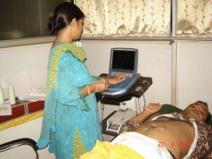 |
Ultrasound |
|
Ultrasound imaging, also called ultrasound scanning or sonography, involves exposing part of the body to high-frequency sound waves to produce pictures of the inside of the body. Ultrasound exams do not use ionizing radiation (as used in x-rays). Because ultrasound images are captured in real-time, they can show the structure and movement of the body's internal organs, as well as blood flowing through blood vessels. Ultrasound imaging is a noninvasive medical test that helps physicians diagnose and treat medical conditions. |
| |
|
| |
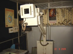 |
Ultrasound |
|
X-rays are a form of electromagnetic radiation, just like visible light. In a health care setting, a machines sends are individual x-ray particles, called photons. These particles pass through the body. A computer or special film is used to record the images that are created. Structures that are dense (such as bone) will block most of the x-ray particles, and will appear white. Metal and contrast media (special dye used to highlight areas of the body) will also appear white. Structures containing air will be black and muscle, fat, and fluid will appear as shades of gray. |
|
